In this session we will build the setup for the KcsA, a membrane protein, and the step to perform the simulation1.
The files you need are available here.
Disclaimer
EDITED: ee will use CHARMm 36 ff.
You are, as always, encourage to read the tutorial in the Notes, taking into account that the forcefield used in the referenced tutorial is CHARMm27.
Membrane protein
Membrane proteins are usually more difficult to simulate with respect to soluble proteins. The difficulties are mainly related to the unavailability to have experimental crystal structures, and to the longer timescale that must be explored.
We will build the setup of the KcsA, a potassium channel membrane protein.
Your setup will look like this:
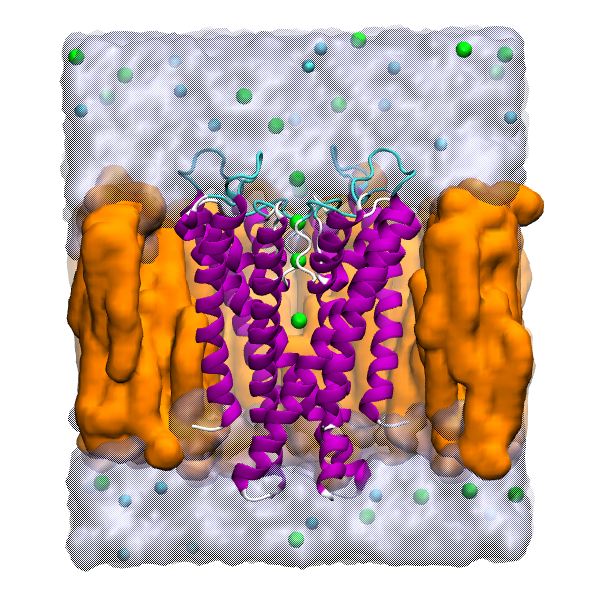
Except from the change in the ion composition (this time KCL), we can see that obviously we need to add a membrane patch.
Our challenge
If we over-simplify the steps, our goal is to apply the following equation.
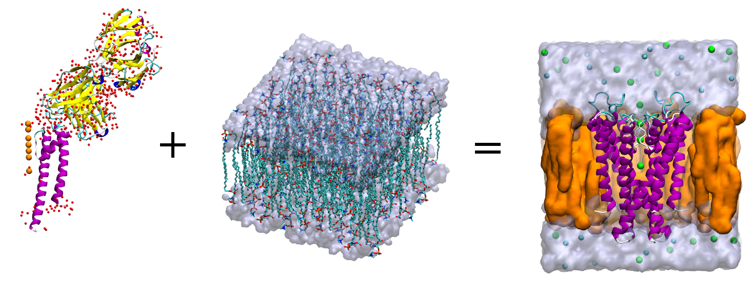
The equation of love <3.
How do we go from the left side of the equation to the right side?
The road map is:
- Download and modify the experimental
pdb; - Obtain the tetramer;
- Create a membrane patch;
- Insert the protein inside the membrane;
- Solvate and ionise the sytem.
Let’s see in details all the steps.
The protein
Download from the PDB website the entry 1K4C.
Have a look at the biological assembly!
From the website you can already see how the protein is arranged within the membrane.
Let’s open the pdb in VMD and visualise it.
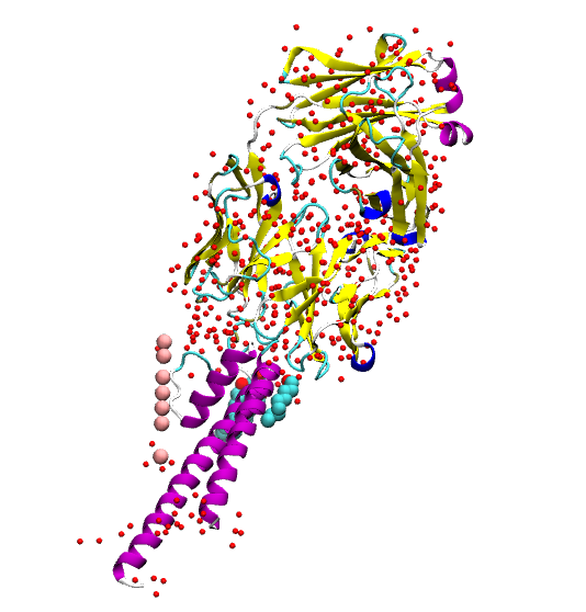
In this system we see that there is not only our protein (the alpha helices). We have also water molecules, several potassium ions, few lipids and antibody fragments that is used to crystalise the protein.
Visualise the protein secondary structure and colour it by chain.
How many chain are there?
Take note of the chain we need for our setup.
Let’s now investigate the pdb file, and go and check the remarks 350,
465 and 470.
What do these remarks tell us?
Now we have two pieces of information: the first one is that our system is a
tetramer (REMARKS 350); the second one is that we have the matrix to apply
to the system to obtain the biological assembly.
As you can see the matrix is a 3 by 4 matrix, to which we have to add a
row {0.0 0.0 0.0 1.0} to obtain a 4 by 4 matrix that is used by VMD
to apply roto-translations.
Let’s start from building the whole tetramer. The axis of rotation will be the z axis,
and the tetramer will be built arount the K ions.
What we are doing here is to create 4 pdb files with a different segname.
After loading the 1K4C.pdb, let’s set an appropriate segname for the first
element of the tetramer (to which we will apply the identity matrix). So we just
set the segname only.
mol new 1K4C.pdb
set all [atomselect top all]
$all set segname A
$all writepdb KCSA-A.pdb
$all delete
Then we apply the second matrix to our system and we change the segname too.
Of course we save the system also this time.
Check the new selection!
set sel [atomselect top "all and not name K"]
$sel set segname B
$sel move { {-1.0 0.0 0.0 310.66} {0.0 -1.0 0.0 310.66} {0.0 0.0 1.0 0.0} {0.0 0.0 0.0 1.0} }
$sel writepdb KCSA-B.pdb
$sel delete
mol delete top
At the end we deleted the structure, since we changed the coordinates and the matrices are given for the given pdb.
Complete the tetramer by applying the other transformations.
When you have the four files, we need to join them in one file:
$ cat KCSA-A.pdb KCSA-B.pdb KCSA-C.pdb KCSA-D.pdb > KCSA.pdb
You have to delete the lines END and CRYST1 ... that you have in the
middle of your file.
Visualise the whole system
The system is now complete, but we want to simulate only the channel, therefore we need to emend the structure.
Let’s remind what we are doing.
The goal is to create the psf for the tetramer. To do so, we need a
psf for each part of the system that is not covalently bonded to other parts.
First, let’s save only the part we are interested in. Of course we will have several pdb files, one for each segment.
Due to the definition of the segname in the previous step,
we can introduce a new for loop in Tcl that iterates over all the
elements of a list: foreach.
Check the script before you execute them, since there are missing values to be filled in!
Load the KCSA.pdb and in the TkConsole:
foreach S { A B C D } {
  set seg [atomselect top "segname $S and chain _whatChain?_ and protein"]
  $seg writepdb seg$S.pdb
  $seg delete
}
In this way, we wrote a pdb file for each monomer.
Check the new files!
We want also to save the potassium ions. We will save only four out of the
seven atoms.

We selected two positions of the ions that are known to be occupied in the filter, and we keep also two ions, one in the extracellular and one in the intracellular region.
Pay attention to the 'atom name' of the potassium atoms!
set pot [atomselect top "name K and resid 3001 3003 3005 3006"]
$pot set name POT
$pot set resname POT
$pot writepdb pot.pdb
We will modify the remaining three K into water molecules.
It’s a kind of magic
set npot [atomselect top "name K and resid 3002 3004 3007"]
$npot set name OH2
$npot set resname TIP3
$npot writepdb filtwat.pdb
We have now two more files: pot.pdb and filtwat.pdb
We want also to save the water crystalised nearby the channel:
foreach S {A B C D} {
  set wat [atomselect top "segname $S and
  resname HOH and within 10 of (chain C and protein)"]
  $wat writepdb crystwat$S.pdb
  $wat delete
}
Now the system is almost ready for the creation of the psf file.
Before that, we need to check the protonation states of charged residue and
of the hystidines.
First, visualise in the KCSA.pdb the hystidine and cysteines.
Are there disulfide bonds? (Check the distances)
The hystidine will be set as HSE.
For the charged residue, visualise in Licorice charged and chain C.
Most of the arginines are located in regions likely to be solvated, so they
are kept charged.
Take a closer look at ASP80 and GLU71. They are both negatively charged,
so problably one of them is protonated (they interact also with a water
molecule). We will define the glutamic acid as GLUP (it’s a patch!).
We are now ready to create the psf.
Write a file kcsa_psfgen.tcl:
mol delete all
package require psfgen
topology _fill-me-prot_
topology _fill-me-wat_
pdbalias residue HIS HSE
pdbalias atom ILE CD1 CD
pdbalias atom HOH O OH2
pdbalias residue HOH TIP3
foreach S { A B C D } {
  segment $S {
    pdb seg$S.pdb
  }
  coordpdb seg$S.pdb $S
  patch GLUP $S:71
  regenerate angles dihedrals

  segment WC$S {
    auto none
    pdb crystwat$S.pdb
  }
  coordpdb crystwat$S.pdb WC$S
}

segment I {
  pdb pot.pdb
}
coordpdb pot.pdb I

segment WF {
  auto none
  pdb filtwat.pdb
}

coordpdb filtwat.pdb WF
guesscoord
writepdb kcsav.pdb
writepsf kcsav.psf
Launch the script and check if there are errors in the output.
Solvate (optional)
We would like to add a water shell to our protein. To do so, we can you a software by the Grubmueller’s group. You have to download solvate and, after compiling it, you should run:
solvate -t 3 -n 8 -w kcsav solkcsa
and then you should join the protein and the water shell. Here there is the script you should use, but the complete files are already provided.
set sol_infile solkcsa.pdb
set kcsa_inbase kcsav
set outbase kcsav_solv_raw
package require psfgen
resetpsf
topology _fill-me-with-water-top_
segment SOLV {
  auto none
  first NONE
  last NONE
  pdb $sol infile
}
coordpdb $sol_infile SOLV
readpsf ${kcsa_inbase}.psf
coordpdb ${kcsa_inbase}.pdb
writepdb ${outbase}.pdb
writepsf ${outbase}.psf
Protein orientation
The goal of this subsection is to orient correctly the protein.
First, load the psf and pdb file and let’s center our system.
set all [atomselect top "all"]
$all moveby [vecinvert [measure center $all]]
display resetview
The membrane patch will have a thickness of 30 Angstrom. We will delete the water molecules that are in hydrophobic region of the protein.
Visualise the protein in VdW and ResType as Coloring Method.
We will also colour the water molecules according to their z coordinates.
Let’s colour the water in Blue. Add two more representations for the
water and z < -20 and water and z>10 in Red. You should have
something like:
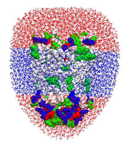
Always visualise that you are going to remove!
In order to remove the central water, the scheme to be followed is:
- define a selection that contains the molecules we want to get rid of;
- assign to the selection a custom
betafactor (usually1); - extract a
segidandresidfor each molecule withbeta > 1(for water we will select only the oxygen atom); - load
psfandpdband delete the unwanted residues.
In practice, we select only the water added later with the solvate
external package:
set solv [atomselect top "segname SOLV"]
$solv set beta 1
Then we set beta 0 to the water molecule we want to retain.
set seltext "segname SOLV and same residue as ((z < -20) or (z>10))"
set sel [atomselect top $seltext]
$sel set beta 0
and then we build two lists with segid and resid that univocally define
a residue.
set badwater [atomselect top "name OH2 and beta > 0"]
set seglist [$badwater get segid]
set reslist [$badwater get resid]
Finally, we delete the select residues.
mol delete all
package require psfgen
resetpsf
topology _path_to_top all27 prot lipid.rtf
readpsf kcsav_solv_raw.psf
coordpdb kcsav_solv_raw.pdb
foreach segid $seglist resid $reslist {
delatom $segid $resid
}
writepdb kcsa_solv.pdb
writepsf kcsa_solv.psf
The position of the protein inside a membrane can be identified visually by the hydrophobic region of the protein, usually limited by rings of Tyrosine. There are available also web software, like OPM, that gives a plausible orientation for membrane proteins.
Membrane patch
Now we want to model a membrane.
Create a membrane 90x90 Angstrom of POPC (a kind of lipids generally used im membrane protein simulation). Use the CHARMm 27 ff.
Find the way to build the membrane
After building it, the membrane, oriented with the z axis normal to its surface, is loaded by default.
Center the membrane and same the new coords as popc_TEMP.pdb
As a convention, the setup has the membrane parallel to the xy plane, and the protein is aligned to the z axis.
Load the kcsa_solv.p*. We want to center the protein using the center of mass
of residue 97 to 106.
set vest [atomselect top "protein and resid 97 to 106"]
$kcsa moveby [vecinvert [measure center $vest weight mass]]
display resetview
$kcsa move [transaxis z -25]
$kcsa writepdb kcsa_TEMP.pdb
And we now join the two structure: protein and membrane.
mol delete all
package require psfgen
resetpsf
readpsf popc.psf
coordpdb popc_TEMP.pdb
readpsf kcsa_solv.psf
coordpdb kcsa_TEMP.pdb
writepsf kcsa_popc_raw.psf
writepdb kcsa_popc_raw.pdb
file delete kcsa_TEMP.pdb
file delete popc_TEMP.pdb
Notes
-
See Membrane tutorial for the references. ↩